Custom Search

Causes of Bilateral diminished concentration of contrast material in the pelvocalyceal system

1- Overhydration/ inadequate dehydration :
Causes dilution of the contrast material (Note that the kidneys may be entirely normal).

2- Polyuria :
Excretion of large volumes of hypotonic urine due to diuretic therapy, diabetes insipidus (lack of antidiuretic hormone ADH secreted by the posterior lobe of the pituitary gland), diabetes mellitus, and intrinsic renal diseases.

3- Renal failure (uremia) :
Severely decreased renal function due to a variety of underlying causes.

4- Nephrosclerosis :
Long-standing hypertension causes narrowing of extraand intrarenal arteries with prolonged intrarenal circulation time and decreased excretion of contrast material.
Malignant nephrosclerosis. Nephrotomogram obtained 5 minutes after the injection of contrast material shows minimum opacification of small, smooth kidneys.

5- Technical :
Injection of an inadequate dose of contrast material.


Causes of Bilateral diminished concentration of contrast material in the pelvocalyceal system


Lymphoma in the Brain of transplant recipient paient

Single or multiple ring-enhancing lesions that primarily affect transplant recipients (high incidence of central nervous system lymphoma in these patients).

Lymphoma developing after renal transplantation. Heart-shaped, peripherally enhancing, central lucent lesion (arrow) situated in the frontoparietal region. There is moderate surrounding edema.

A case of acute Epiglottitis in an Adult Leukemia Patient

Epiglottitis is defined as an acute infection of the supraglottic larynx which may require an emergency airway management.
In Emergency room you can diagnose a patient by symptoms of airway obstruction. Fibreoptic laryngosocpy (see the case report) or soft tissue lateral x-ray neck (see picture below) are the other preferred methods of diagnosis. Indirect laryngoscopy should be done with caution since patient gagging may worsen or participate catastrophic airway obstruction.
This case report describes a neutropenic patient with epiglottitis who was subsequently diagnosed as having acute promyelocytic leukemia.
Case Report: A 50 year old woman presented with a week long history of pain on swallowing and dysphagia. The odynophagia was gradually progressive and more for solids than liquids.
4 months back, she had been diagnosed as having tuberculous lymphadenitis and started on anti koch's treatment (AKT). The AKT had to be stopped three days before she came to us because of persistent neutropenia.
On examination, she was otherwise normal with no temperature or dyspnea. On palpation, she did have a slight tenderness of the laryngeal frame work. We were unable to find a definitive cause of pain in the throat and performed an indirect laryngoscopic examination which revealed a ‘cherry-red’ epiglottitis with slough at the base of the epiglottis. Fibreoptic laryngoscopy confirmed the diagnosis of epiglottitis (As seen in the picture ).
She was admitted for observation and emergency airway management if such a situation arose. A throat swab was sent and routine haemogram(CBC) started her on Ceftriaxone. The doctor did not give her steroids as she was a diabetic and was also immunosupressed.
Her haemogram was repeated daily and it showed a persistent low white blood cell count (WBC) and platelet count. The throat swab revealed Beta hemolytic Streptococcus pyogenes sensitive to Cephalosporins. When her CBC did not improve even after three days of administering granulocyte-macrophage colony stimulating factor, A bone marrow biopsy was done. This revealed that she was suffering from acute promyelocytic leukemia with thrombocytopenia .

She was started on induction chemotherapy, supported by multiple blood and blood factor transfusions.  The disease went into remission on the 18th day

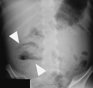
Ruptured Appedicitis in abdominal X-ray

Ruptured appendicitis (arrow heads).


Note the presence of an ill-defined mottled gas pattern within the RIF,with an air–fluid level, caused by the ruptured appendix.

Role of Abdominal X-ray in Appendicitis

The aetiology of Appendicitis is usually related to luminal obstruction,often by lymphoid hyperplasia or a faecolith.

Radiological features in abdominal X-ray:
• Abdominal X-ray Is neither sensitive nor specific for Appendicitis but can provide clues.
• The presence of a calcified appendicolith in the RLQ ,combined with
abdominal pain,has ahigh positive predictive value for acute appendicitis.
• Other signs are less specific and include caecal wall thickening,small-bowel ileus and decreased small-bowel gas in the RIF.
• Free peritoneal fluid can lead to loss of the psoas out line,loss of the fat planes around the bladder and loss of definition of the inferior liver outline.

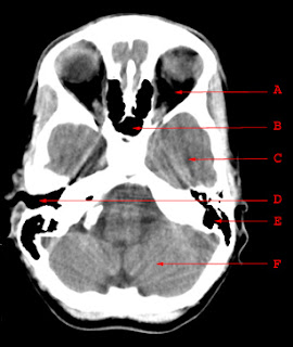
Normal Anatomy of CT brain at skull base


A. Orbit

B. Sphenoid Sinus

C. Temporal Lobe

D. External Auditory Canal

E. Mastoid Air Cells

F. Cerebellar Hemisphere

Asbestos related pleural plaques

Asbestos plaques appeare as Calcified asbestos related pleural plaques and they have a characteristic appearance, and are generally considered to be benign.
They are irregular, well-defined, and classically said to look like holly leaves.

Asbestos related pleural plaques

Temporomandibular Joints (TMJ) Radiographic Anatomy

Adult Lateral Oblique - Closed Mouth


Adult Lateral Oblique - Open Mouth

 Localized osteoporosis due to Burn, Frostbite and Electric shock

The bone demineralization is most marked where softtissue damage was greatest, is an early radiographic finding that may persist for a prolonged period.

Electrical injury. This X-ray shows comminuted fracture of the head and shaft of the humerus associated with mottled decalcification of the humeral head.
The cortex of the humerus is thin, and the medullary cavity is widened. Discrete areas of rarefaction can be seen in the shaft and distal metaphyseal region.

Sonography of fibroid with calcification


This middle aged female patient complained of pain in the suprapubic region. Ultrasound imaging revealed a small uterus with a calcific well-defined, intramural mass in the lower part of the body of uterus.
There is clear acoustic shadowing posterior to the calcific lesion. These findings suggest a calcific fibroid of the uterus. Fibroids are often known to undergo calcificaiton in elderly females.

Disuse osteoporosis follows prolonged immobilization

To maintain osteoblastic activity at normal levels, bones must be subjected to a normal amount of stress and muscular activity. Within a few weeks after the fracture of a bone, localized osteoporosis becomes detectable, especially distal to the site of injury.

The cortical margin of an involved bone never completely disappears (unlike bone destruction due to disease). Similar disuse atrophy due to immobilization follows neural or muscular paralysis.


Disuse osteoporosis. (A) Severe periarticular demineralization follows prolonged immobilization of the extremity.


(B) In a patient with a fractured patella, there is pronounced subcortical demineralization in the distal femur. The cortical margin (arrows) remains intact.

Ischaemic colitis Clinical and Radiological findings

Clinical characteristics
• Ischaemic colitis is caused by interruption to the colonic blood supply that include thrombosis , bowel obstruction and trauma.
• Some Predisposing factors included as age,oral contraceptives,sickle cell disease and surgical ligation of the inferior mesenteric artery.
• Presents with acute lower abdominal pain and tenderness,usually out of proportion to the clinical signs.There may be rectal bleeding or diarrhoea.
• Most commonly affects the left side of the colon, especially at the splenic flexure where there is a watershed between the territories of the superior and inferior mesenteric arteries.The rectum is usually spared.
• May be a transient condition with spontaneous resolution over a few months. May lead to incomplete healing with smooth stricture formation. Severe disease can lead to colonic infarction,with a high associated mortality.

Radiological features
• AXR : plain film is often normal;however,gas within the colon may out line the characteristic thumb printing of thickened, oedematous folds seen in this condition.
• Barium enema : single-contrast instant enema may demonstrate thumb printing and ulceration associated with this condition.Adoublecontrast enema shows these findings more reliably but should be used with caution in anacutely ill patient.A smooth stricture maybe demonstrated on a delayed study.
Late-stage ischaemic colitis. Single-contrast barium enema demonstrates a clear zone of transition between normal and abnormal colon at the junction of the middle and distal thirds of the transverse colon.The proximal colon has normal mucosa and haustral pattern while the distal
segment is featureless and abnormally narrowed.
• CT :contrast-enhanced spiral CT is the usual first-line investigation for suspected ischaemic colitis.Adual phase scan,performed in the arterial and portal phases,may demonstrate thrombus in both the mesenteric arterial or venous systems.The affected colon may appear abnormally circumferentially thickened and demonstrate poor contrast enhancement.There may be a sharp cut off between normal and abnormal colon at the boundary of vascular territories.Mural gas may be seen in more advanced disease and,in severe cases,portal gas may be identified. The latter is a poor prognostic factor.
Superior mesenteric artery (SMA) thrombosis. Normal enhancement of the aorta (arrowhead). No enhancement seen in the SMA (arrow).
• Angiography :a more limited role in the era of multislice spiral CT but may demonstrate attenuated arterial flow or the presence of a thrombus.


Ischaemic colitis Clinical and Radiological findings


Associations of Absent thumb cases

An absent thumb can have many associations. They include :

* Fanconi anemia (pancytopenia-dysmelia syndrome)
* Franceschetti syndrome
* Holt-Oram syndrome
* phocomelia (e.g. thalidomide embryopathy)
* Poland syndrome (pectoral muscle aplasia - syndactyly)
* Rothmund-Thomson syndrome
* Seckel syndrome (bird-headed dwarfism)
* trisomy 18
* Yunis-VarĂ³n syndrome

Rickets X-ray Before and After vitamin D therapy

Systemic disease of infancy and childhood in which calcification of growing skeletal elements is defective because of a deficiency of vitamin D in the diet or a lack of exposure to ultraviolet radiation (sunshine).
Most common in premature infants and usually develops between 6 and 12 months of age.

Classic radiographic signs :
It include cupping and fraying of metaphyseal ends of bone with disappearance of normally sharp metaphyseal lines; delayed appearance of epiphyseal ossification centers, which have blurred margins (unlike the sharp outlines in scurvy); and excessive osteoid tissue in the sternal ends of ribs producing characteristic beading (rachitic rosary).

(A) Initial film shows severe metaphyseal changes involving the distal femurs and proximal tibias and fibulas. Note the pronounced demineralization of the epiphyseal ossification centers.


(B) After vitamin D therapy, there is remineralization of the metaphyses and an almost normal appearance of the epiphyseal ossification centers.

What is Ultrasound Gain–correct, over, under (with photos)

Gain refers to the control sonologists use to adjust the brightness with which returning sound waves are displayed by the ultrasound machine. When an echo returns from body tissue, it does so within an amplitude range.
The ultrasound device translates that amplitude range to a brightness, which it displays on the monitor. The overall gain allows the sonologist to adjust the brightness of all returning echoes. Decreasing the gain makes the overall image less bright, while increasing the gain makes the image more bright .

These figure shows the types of gain:

(A) (Correct) This image is correctly gained

(B) (Over) The image has too much gain applied to the image. Compared with image A, echoes are found where there should be none.

(C) (Under) The image is undergained. The image is too dark, potentially making it difficult for accurate diagnosis.

Syphilitic Aortitis in Chest X-ray

Syphilitic Aortitis in Chest X-ray appears as a dilatation of the ascending aorta, frequently with mural calcification

It may cause inflammation of the aortic valvular ring that results in aortic insufficiency. Approximately one-third of patients develop narrowing of the coronary ostia that may lead to symptoms of ischemic heart disease.

Syphilitic aortitis. Aneurysmal dilatation of the ascending aorta with extensive linear calcification of the wall (arrows). Some calcification is also seen in the distal aortic arch.

Madelung's Disease (benign symmetric lipomatosis)

A 45-year-old man presented with a 3-year history of a painless, soft, and slow-growing swelling of the neck, upper trunk, upper back, and shoulders (Panels A and B). The patient had a history of heavy alcohol consumption and was a nonsmoker. Laboratory blood analysis showed minor elevations in aspartate aminotransferase (71 U per liter), alanine aminotransferase (49 U per liter), and total cholesterol (235 mg per deciliter [6.08 mmol per liter]). Triglycerides were very elevated at 1020 mg per deciliter (11.52 mmol per liter).
Magnetic resonance imaging revealed diffuse, nonencapsulated fatty deposits in the mediastinum and in the subcutaneous and deeper fascial compartments of the neck, upper trunk, and back (Panel C, arrows). A clinical diagnosis of Madelung's disease was made.

Madelung's disease (also known as benign symmetric lipomatosis, the Launois–Bensaude syndrome, and multiple symmetric lipomatosis) is a rare disorder of unknown cause. In reported case series, up to 90% of patients have a history of chronic alcoholism, and there is a strong male predominance. Since the patient was asymptomatic, no surgical treatment was proposed. He was started on lipid-lowering therapy and referred to an alcohol detoxification program.

Frog leg x-ray view in slipped capital femoral epiphysis

What is Frog leg x-ray view ??
frogleg lateral view or may be called "cossak dancer`s view" (abducted lateral) is obtained when patient is supine with knees flexed, soles of feet together, and the thighs maximally abducted;the central beam is directed vertically or with a 10 to 15 deg cephalic tilt to a point slightly above pubic symphysis.

OK;
But what is slipped capital femoral epiphysis (SCFE) ??
Slipped capital femoral epiphysis (SCFE) or may be also called "Slipped upper femoral epiphysis" is a fracture through the physis (the growth plate), which results in slippage of the overlying epiphysis.And any abnormal movement along the growth plate results in the slip .
It is a common cause of hip and knee pain in children aged 10–17 that occures in adolescents ,often overweight children, especially boys.

X-ray showing a slipped capital femoral epiphysis, before and after surgical fixation.

Note that :Early on in the disease process the femoral metaphysis shifts anterosuperior relative to the epiphysis. AP views of the hip may be normal initially with slippage first noted on the frog leg (lateral) view.

Crescent sign of femoral AVN

Definition :
The crescent sign that is associated with avascular necrosis (AVN) is seen on conventional radiographic films and is recognized as a curvilinear subchondral radiolucent line . It is typically seen along the anterolateral aspect of the proximal femoral head, which is optimally depicted on the frog-leg radiographic view "obtained with the patient’s thigh abducted and flexed ".

A Conventional radiograph of the right femur in the frog-leg position shows subchondral area of hyperlucency (arrows) in the anterolateral aspect of the proximal femoral head. (Courtesy of Clyde A. Helms, MD, Department of Radiology, Duke University Medical Center, Durham, NC.)


EXPLANATION
The crescent sign is explaned by Inadequate perfusion in the articular ends of bones that leads to the processes of osteonecrosis and repair . Repair begins at the interface between necrotic and viable bone.
Reactive new bone is laid down over dead trabeculae, which produces a sclerotic margin. An advancing front of fibrosis, hyperemia, inflammation, and bone resorption extends into the necrotic segment of bone as repair is attempted. Mechanical failure of trabecular bone at this interface results in progressive microfracture (as seen below ) and collapse of the adjacent dead subchondral cancellous trabeculae, which leads to the development of a subchondral radiolucent area along the fracture line, or the crescent sign
Specimen radiograph of a coronally sectioned femoral head segment reveals a subchondral fracture (arrows), which manifests as the crescent sign. Note the fragmentation and compaction of the subchondral cancellous trabeculae, which weakens the articulating surface.

A brief introduction to Acute Vs Chronic Subdural haematoma in CT

Subdural haematomas arise between the dura and arachnoid, usually from ruptured veins crossing this potential space. The space enlarges as the brain atrophies and so subdural haematomas are more common in the elderly.
There are 2 types of subdural haematomas :
Acute subdural - Chronic subdural

Acute subdural
It is presents in a similar fashion to the extradural haematoma, and can have equally severe consequences due to mass effect, requiring urgent surgery. So Differentiating the two is not so important in the acute situation.

The blood is again of high attenuation, but may spread more widely in the subdural space, with a crescentic appearance and a more irregular inner margin.

Chronic subdural
It`s aetiology is not always clear. It is probably due to trauma, often minor, in the preceding few weeks, but no such history is obtainable in 50% of cases. Symptoms are vague and often develop slowly with a gradual depression or fluctuation of conciousness. In 10% of cases subdural haematomas are bilateral .

While acute subdural haematomas have increased attenuation, this decreases with time, becoming isodense after a week or so, and hypodense thereafter. Consequently chronic subdurals are often hypodense crescentic collections, often with mass effect. The collection may be more complex with layering of more dense material posteriorly and a gradual transition. Expansion due to osmosis may tear further veins leading to recurrent bleeds; hyperdense red blood cells from fresh bleeding may layer posteriorly, and complex septated collections may develop.

Isodense collections may be better demonstrated after intravenous contrast as the density will then be less than that of the brain. However this is rarely a problem with more modern scanners.

Basic Gross anatomy of the human heart

A brief introduction to Extradural haematoma in CT

Extradural haematoma arises between the inner table of the skull and the dura of the brain. They usually develop from injury to the middle meningeal artery or one of its branches,So it is usually temporoparietal in location.
A temporal bone fracture is often the cause, but is not essential. The expanding haematoma separates the dura from the skull; this attachment is quite strong such that the haematoma is confined, giving rise to its characteristic biconvex shape, with a well defined margin.

It may present as primary depressed consciousness or following a lucid interval. The bleeding is usually acute and so of high attenuation. There is often significant mass effect with compression of the ipslateral lateral ventricle and dilatation of the opposite lateral ventricle due to obstruction of the foramen of Munro. The basal cisterns may be effaced.

This is the typical appearance and location of an acute extradural haematoma. Note the high density of the haematoma and slight midline shift .

3D Virtual Colonoscopy

3D Virtual Colonoscopy at Associated Radiologist Warren NJ, Edison NJ, and Bridgewater NJ offices.

Coiled-spring appearance of Intussusception

Intestinal Intussusception in abdominal x-ray may produce the classic coiled-spring appearance (barium trapped between the intussusceptum and the surrounding portions of bowel).

Note that Intestinal Intussusception is a major cause of small bowel obstruction in children (much less common in adults). The leading edge of an intussusception (usually a pedunculated polypoid tumor) can be demonstrated in 80% of adults. In children, there is infrequently any apparent anatomic etiology.

Intussusception. Coiled-spring appearance (arrow) in jejuno-jejunal intussusception.

X-ray of Gallstone ileus

Imaging Findings :

Classic triad of
  1. jejunal or ileal filling defect
  2. gas or barium in the biliary tree
  3. small bowel obstruction.

Gallstone ileus is caused by a large gallstone entering the small bowel via a fistula from the gallbladder or the common bile duct to the duodenum. Usually occurs in elderly women.

Gallstone ileus. Upper gastrointestinal series demonstrates the obstructing stone (white arrows) and barium in the biliary tree (black arrow).