Custom Search

Contraindications to MRI procedure

The principal contraindications of the MRI procedure are mostly related to the presence of metallic implants in a patient. The risks of MRI scans increase with the used field strength. In general, implants are becoming increasingly MR safe and an individual evaluation is carried out for each case.

Absolute Contraindications for the MRI scan:
# Intracranial aneurysm clips (Unless the referring physician is certain that it is made of nonferromagnetic material such as titanium).

# Intra-orbital metal fragments

# Any electrically, magnetically or mechanically activated implants (including cardiac pacemakers, biostimulators, neurostimulators, cochlear implants, and hearing aids).


Relative Contraindications for the MRI scan:
-cochlear implants
-other pacemakers, e.g. for the carotid sinus
-insulin pumps and nerve stimulators
-lead wires or similar wires (MRI Safety risk)
-prosthetic heart valves (in high fields, if dehiscence is suspected)
-haemostatic clips (body)
-non-ferromagnetic stapedial implants

Normal Anatomy of Gall bladder


This ultrasound images show the normal anatomy of the gall bladder taken from different angles of the transducer during sonography. The parts seen here are the fundus, body, neck and the cystic duct.
Note the close relation of the cystic duct to the portal vein. The cystic duct is well seen and appears tortuous in these images.

Obstructive hydrocephalus at Level of foramen of Monro

In CT scan you can find Enlargement of the lateral ventricles with normalsized third and fourth ventricles.

Causes: Colloid cyst; suprasellar tumors (especially craniopharyngioma); intraventricular tumors; arachnoid cysts of the suprasellar cistern; intraventricular hemorrhage (trauma, arteriovenous malformation, hemophilia). Unilateral tumors, such as those arising in the hypothalamus, basal ganglia, or cerebral parenchyma, may obstruct only one side and cause dilatation of the opposite ventricle and mass compression of the ipsilateral ventricle.

Level of the foramen of Monro. (A) Bilateral enlargement of the frontal horns with a normal-sized third ventricle in a patient with a hyperdense colloid cyst (c). (B) Unilateral enlargement of the left frontal horn caused by a tiny hypodense unilateral tumor (arrow).

Happy Holidays from Stanford Hospital & Clinics 2010

Stanford Hospital & Clinics asked its employees what they are thankful for this holiday season, here are their answers.

Stanford Hospital & Clinics wishes everyone a very happy hoiday season and a happy New Year!

Skull (Anterior, Lateral & Mandible) Bony Landmarks

About Technique of Head CT

In order to perform a head CT, the patient is placed on the CT table in a supine position and the tube rotates around the patient in the gantry. In order to prevent unnecessary irradiation of the orbits and especially the lenses, Head CTs are performed at an angle parallel to the base of the skull. Slice thickness may vary, but in general, it is between 5 and 10 mm for a routine Head CT. Intravenous contrast is not routinely used, but may be useful for evaluation of tumors, cerebral infections, and in some cases for the evaluation of stroke patients.

Differences between sutures and fractures in skull x-ray

Linear fracture results from low-energy blunt trauma over a wide surface area of the skull. It runs through the entire thickness of the bone and, by itself, is of little significance except when it runs through a vascular channel, venous sinus groove, or a suture.
In these situations, it may cause epidural hematoma, venous sinus thrombosis and occlusion, and sutural diastasis, respectively. Differences between sutures and fractures are summarized in this Table
Click here for enlargment

Brain Stem Model - Sagittal View - Thalamus & Epithalamus

Pneumonia X-ray

The patient shown below most likely has:


a. A large left pleural effusion

b.
A large right pneumothorax

c.
Atelectasis of the left lung

d.
Pneumonia in the left lung

e.
Unilateral pulmonary edema

The correct answer :
Pneumonia in the left lung

Explanation
-----------
There is opacification of the left hemithorax. There is no shift of the heart and mediastinal structures (i.e. the trachea). This eliminates pleural effusion and atelectasis as possibilities. A tension pneumothorax would displace the heart and mediastinal structures away from the side of the pneumothorax and would not cause complete opacification of the opposite lung. In addition, there should be no lung markings visible in the hemithorax which contains the pneumothorax. Either pneumonia or pulmonary edema could cause the findings shown, but pneumonia is a much more common cause.


Tethered cord syndrome

Clinically presents with motor and sensory dysfunction of the lower extremities (unrelated to myotomal or dermatomal pattern), muscle atrophy, decreased or hyperactive reflexes, urinary incontinence, spastic gait, scoliosis, or foot deformities.
Causes include lipomatous lesions (intramedullary lipomas, lipomyelomeningoceles, lipoma of the filum terminale); myelomeningocele; diastematomyelia; and a short, thickened filum terminale (>2 mm).

Imaging Findings :
Caudal displacement of the conus below the L2L3 level in neonates and young children or below the middle of L2 after age 12.
Tethered cord syndrome. Sagittal T1-weighted image in a patient who had undergone a myelomeningocele repair at birth shows that the cord ends at the L5 level (straight arrow). Note the absence of the posterior elements of the sacrum, as well as the presence of a high-signal-intensity mass (lipoma) within the sacral spinal canal (curved arrows).

Normal Anatomy of Lateral meniscus in MRI

On sagittal images the posterior horn is higher in position than the anterior horn.
Both horns are about the same size.

 The lateral meniscus posteriorly comes up higher over the tibial spine to insert near the posterior cruciate ligament.
This upward position of the posterior horn may be the reason for the higher signal intensity of the posterior horn in all planes due to magic angle effect.
Lateral meniscus: posterior horn and posterior meniscal root.

Normal Anatomy of Medial meniscus in MRI

Both horns are triangular in shape and have very sharp points.
The posterior horn is always larger than the anterior horn (figure).
If this is not the case than the shape is abnormal, which can be a sign of a meniscal tear or a partial meniscectomy.
Medial meniscus: The posterior horn is always larger than the anterior horn.

The posterior root is immediately anterior to the posterior cruciate ligament.
If it is missing on the sagittal images, then there is a meniscal root tear (figure).
The anterior horn has an insertion on the tibia and a second portion that travels from medial to lateral to connect to the anterior horn of the lateral meniscus ( intermeniscal or transverse ligament).
LEFT: normal medial meniscal root immediately anterior to the posterior cruciate ligament.
RIGHT: missing posterior root due to meniscal root tear.

Peptic ulcer disease

If the ulcer crater is very shallow, a thin layer of barium coating results in a ring shadow. Irregular folds merging into a mound of polypoid tissue around the crater suggest a malignancy. Fundal ulcers above the level of the cardia are usually malignant.

Imaging Findings :
Classic signs of benignancy include penetration, Hampton line, ulcer collar, ulcer mound, and radiation of smooth, slender mucosal folds to the edge of the crater.

Fold patterns in gastric ulcers (arrow). (A) Small, slender folds radiating to the edge of a benign ulcer. (B) Thick folds radiating to an irregular mound of tissue surrounding a malignant gastric ulcer (arrow).
Computed tomography in a 52-year-old woman with mild episodic gross hematuria revealed compression of the left renal vein between the superior mesenteric artery (SMA) and the aorta (Ao) before the vein merges into the inferior vena cava (IVC).

This compression caused marked dilatation of the distal part of the renal vein (RV). The renal venous congestion caused hematuria, presumably through the rupture of submucosal veins into the renal pelvis. No treatment was given. Entrapment of the renal vein is a known cause of hematuria.

For more information: HERE

Skull and Face views 2

Image 3:

Findings: Right mandibular condyle fracture
Lesson: Look at the corner of the film

Image 4:

Findings: There is left parietal fracture
Lesson: Do not ignore skull in facial views

Skull and Face views 1

Image 1:

Finding:Fracture body of mandible.
Lesson: Always look the mandible in all facial views


Image 2:

Findings: Orbital emphysema with a tiny soft tissue in the roof of maxillary sinus on the LEFT side, strongly suggestive of orbital blow out (floor of orbit) fracture
Lesson: Look at the soft tissue signs; look for the side marker carefully

Testicular microliths in a new born


The above ulltrasound images and Color Doppler image show testicular microliths (left testis) in a new born baby.
Testicular calcifications can occur in any age group; however the occurrence of this condition in a newborn is very rare. The color Doppler study showed normal vascularity in the affected testis.  
Such cases need to be followed up for changes such as germ cell tumor and more benign conditions such as epididymitis, orchitis and varicocele as well as testicular atrophy. Studies show possible link between Testicular microlithiasis in infants and Down's syndrome.

Caplan Syndrome (Coal worker's)


Posteroanterior radiograph shows a multitude of fairly well-circumscribed nodules and masses ranging in diameter from 1 to 5 cm, scattered randomly throughout both lungs with no notable anatomic predilection. No cavitation is apparent, and there is no evidence of calcification.

This patient, a 56-year-old man, had been a coal miner for many years and in recent years had developed arthralgia, which proved to be due to rheumatoid arthritis. As a means of establishing the nature of the pulmonary nodules, a percutaneous needle aspiration was performed on the large mass situated in the lower portion of the left lung (arrowheads): Several milliliters of inky black fluid were aspirated.

Small-Bowel Intussusception


A previously healthy 22-year-old man presented with a 3-month history of intermittent abdominal pain and hypochromic microcytic anemia, with a hemoglobin level of 5.1 g per deciliter and a mean corpuscular volume of 75 µm3.

Initial endoscopy of the upper and lower gastrointestinal tract was unremarkable, despite a positive test for fecal occult blood. Further investigation with video-capsule enteroscopy showed an intraluminal bulge within the proximal jejunum 77 minutes after ingestion of the capsule. Six days later, the patient presented with abdominal pain, nausea, and vomiting. Computed tomography showed the presence of an intussusception (arrow).

On emergency laparotomy, a proximal jejunojejunal intussusception was found and resected. Pathological examination revealed that the leading edge of the intussusception was a pedunculated benign lipomatous polyp. At a follow-up visit 2 months later, the patient was free of pain and had no further signs of bleeding.

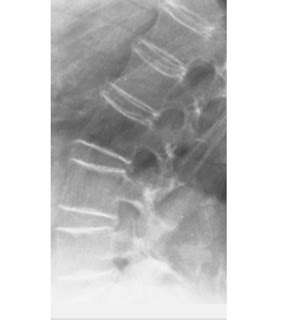
Osteoporosis of aging (senile or postmenopausal osteoporosis)

Most common form of generalized osteoporosis. As a person ages, the bones lose density and become more brittle, fracturing more easily and healing more slowly. Many elderly persons are also less active and have poor diets that are deficient in protein. Females are affected more often and more severely than males, as postmenopausal women have deficient gonadal hormone levels and decreased osteoblastic activity.

 Osteoporosis of aging. Generalized demineralization of the spine in a postmenopausal woman. The cortex appears as a thin line that is relatively dense and prominent (picture-frame pattern).

CT interpretation : pancreas cancer with liver metastasis

Anatomy on Thyroid sonography

The anterior neck is depicted rather well with standard gray scale sonography. (FIGURE 1) The thyroid gland is slightly more echo-dense than the adjacent structures because of its iodine content. It has a homogenous ground glass appearance. Each lobe has a smooth globular-shaped contour and is no more than 3 - 4 centimeters in height, 1 - 1.5 cm in width, and 1 centimeter in depth. The isthmus is identified, anterior to the trachea as a uniform structure that is approximately 0.5 cm in height and 2 - 3 mm in depth.
The pyramidal lobe is not seen unless it is significantly enlarged. In the female, the upper pole of each thyroid lobe may be seen at the level of the thyroid cartilage, lower in the male. The surrounding muscles are of lower echogenicity than the thyroid and tissue planes between muscles are usually identifiable. The air-filled trachea does not transmit the ultrasound and only the anterior portion of the cartilaginous ring is represented by dense, bright echoes. The carotid artery and other blood vessels are echo-free unless they are calcified. The jugular vein is usually in a collapsed condition and it distends with a Valsalva maneuver. There are frequently 1-2 mm echo-free zones on the surface and within the thyroid gland that represent blood vessels. The vascular nature of all of these echoless areas can be demonstrated by color Doppler imaging to differentiate them from cystic structures (10-11).
Lymph nodes may be observed and nerves are generally not seen. The parathyroid glands are observed only when they are enlarged and are less dense ultrasonically than thyroid tissue because of the absence of iodine. The esophagus may be demonstrated behind the medial part of the left thyroid lobe, especially if it is distended by a sip of water. (FIGURE 2)
Figure 1. Sonogram of the neck in the transverse plane showing a normal right thyroid lobe and isthmus. L=small thyroid lobe in a patient who is taking suppressive amounts of L-thyroxine, I=isthmus, T=tracheal ring ( dense white arc is calcification, distal to it is artefact), C=carotid artery ( note the enhanced echoes deep to the fluid-filled blood vessel), J=jugular vein, S=Sternocleidomastoid muscle, m=strap muscle.

Figure 2. Sonogram of the left lobe of the thyroid gland in the transverse plane showing a rounded lobe of a goiter. L=enlarged lobe, I= widened isthmus, T=trachea, C=carotid artery ( note the enhanced echoes deep to the fluid-filled blood vessel), J=jugular vein, S=Sternocleidomastoid muscle, m=strap muscles, E=esophagus.

Complementary ultrasound to mammography ; Why?

Breast cancer can lie hidden from mammography in women with dense breast tissue, and thus the role of ultrasound is implemented to evaluate the texture and anatomical structures within the breast. Women most likely to have dense breasts are younger, premenopausal women and postmenopausal women who are taking hormone replacement therapy.

Breast ultrasound is used as a complementary examination to mammography in the following situations:

* Evaluation of dense breast tissue.
* Evaluation of a mass demonstrated on mammography.
* Guidance of biopsy needle or needle localization (see cyst aspiration image)

Cardiac CT: Basic Principles and Techniques

Grades of hydronephrosis (on Ultrasound imaging):

A) Mild hydronephrosis:
This images shows mild dilatation of the pelvis as well as the calyces of the right kidney suggesting mild hydronephrosis. The left kidney also appears to be affected similarly. This must be differentiated from mild dilatation of the renal pelvis alone (called splitting of the pelvis), which is physiological and transient; this means that on emptying of the urinary bladder, this appearance should disappear.

B) Moderate hydronephrosis:

The above ultrasound images show cupping of the calyces with moderate dilation (Right kidney) of the pelvis and calyces. Despite the hydronephrosis the renal parenchyma is still preserved.

C) Severe hydronephrosis:

The above sonographic images show marked dilatation of the pelvicalyces with sever thinning of the renal parenchyma. note almost total absence of normal renal tissue (cortex).

Pelvi-ureteric junction calculus (Toshiba Nemio-XG ultrasound machine)


This patient had pain in the right lumbar region. ultrasound of the abdomen showed hydronephrosis of the right kidney with a moderately large calculus (14mm.) at the right Pelvi-ureteric junction (arrows). Such calculi cause partial obstruction of the renal pelvis and may cause severe ureteric colic. Ultrasound images of pelvi-ureteric junction calculus, were taken with a Toshiba Nemio-XG ultrasound machine. (Synonyms: PUJ calculus or UPJ calculus or UPJ stones).
Anatomic Consideration : The gastric antrum is generally located posterocaudally to the left lobe of the liver. The proximal stomach is usually difficult to delineate because of the significant artifact arising from gas in the stomach. When filled with liquid contents, the proximal stomach can be identified medially to the splenic hilum with left intercostal or coronal scanning.

In scanning the epigastric region, the gastric antrum can be demonstrated anterior to the pancreatic body and caudal to the left lobe of the liver . Intricate details of the structure of the gastric wall can be demonstrated using a high-frequency probe.
 Normal gastric antrum. (A) In an epigastric sagittal plane, the cross section of antrum (G) is visualized anterior to the pancreatic body (Pb) and caudal to the left lobe of the liver (L). The pancreatic body is located anterior and cephalad to the splenic vein (arrow) and the superior mesenteric artery (SMA). (B) In a transverse plane, the gastric antrum is demonstrated anterior to the pancreatic body. (C) Five-layer structure of the gastric wall is demonstrated in a transverse scanning with a high-frequency probe. Ao = aorta, IVC = inferior vena cava, SV = splenic vein.
Click on photo for enlargment

Mortise view; How to get and What`s represent?

Mortise view : To obtain a better view of the ankle mortise, the patient's leg must be internally rotated just enough so that the lateral malleolus (which is normally posterior to the medial malleolus), is on the same horizontal plane as the medial malleolus, and a line drawn through both malleoli would be parallel to the tabletop.

Usually this only requires approximately 10 - 20 degrees of internal rotation. In other words, when viewing the mortise view, the tibia and fibula must be viewed without superimposition on each other. This mortise view represents a true AP projection of the ankle mortise and also provides a good visualization of the talar dome (to rule-out osteochondral talar dome fractures).

The History of Radiology

Unilateral renal agenesis (solitary kidney)

Imaging Findings:
Filling of the renal fossa with bowel loops (sharply outlined gas or fecal material in the plane of the renal fossa on nephrotomography). The contralateral kidney usually shows compensatory hypertrophy.

NOTES:
Rare anomaly that is associated with a variety of other congenital malformations. It is essential to exclude a nonfunctioning, diseased kidney by ultrasound or CT. After nephrectomy, the renal outline is generally preserved on plain films if the perinephric fat is left in situ.

Echocardiogram

Why does a CT scan cost so much in USA? (billions dollars on Defensive Medicine)


It started as a simple stomach ache, but Alexandra Varipapa, a sophomore at the University of Richmond, decided to go to the emergency room.

There, doctors ordered a full CT scan, a radiation imaging test, which found a harmless ovarian cyst. She never questioned the CT scan, CBS News correspondent Wyatt Andrews reports.

But her father did - when he got the $8,500 bill, $6,500 of which was that CT scan.

“I was pretty flabbergasted,” said Robert Varipapa, himself a physician.

Varipapa says his daughter's pain could have been diagnosed far more easily and cheaply with a $1,400 ultrasound.

“A history, a pelvic examination and probably an ultrasound,” he said. And he would have started with the ultrasound.

But the hospital defends the CT scan, saying an ultrasound might have missed something more serious.

“It would not have ruled out appendicitis obviously, it would not have ruled, necessarily, out a kidney stone,” said Dr. Bob Powell, ER medical director of Bon Secours St. Mary’s Hospital.

Varipapa agrees, but asks why not start simple - and do the CT scan only if necessary?

“Well it's my opinion this is defensive medicine,” Varipapa said.

Defensive medicine is what happens when doctors order too many tests because they are afraid of missing a diagnosis and later losing a multi-million dollar lawsuit for malpractice. Defensive medicine these days is so pervasive, some estimate its yearly cost at more than $100 billion.

Dr. Kevin Pho runs the popular medical blog, Kevin M.D., where doctors routinely confess exactly how they run up costs by practicing defensive medicine.

“Defensive medicine is bad medicine,” Pho said.

In a post, one ER doctor says he's just admitted two patients to the hospital - when he was sure "neither was having cardiac (problems), but what am I to do?"

Another admits that in his practice, “every patient with a headache gets a (CT) scan.”

“It's much easier to defend the fact that you ordered a test than it is to not order the test at all,” Pho said.

And the costs of defensive medicine today are increasingly paid by patients, even those with insurance - because of rising deductibles and co-payments.

“There’s no doubt in my mind this is a significant driver in health care costs today,” Pho said.

Source : CBS News

Esophegeal impression by Thyroid enlargement

Smooth impression on and displacement of the lateral wall of the esophagus, usually with parallel displacement of the trachea.
Caused by localized or generalized hypertrophy of the gland, inflammatory disease, or thyroid malignancy.

Enlargement of the thyroid gland. Smooth impression in the cervical esophagus (arrow).

Ultrasound images of Benign prostatic hyperplasia


This 80 yr. old male patient presented with lower urinary tract symptoms (LUTS). Transabdominal ultrasound scan images reveal obvious intravesical enlargement of the enlarged median lobeof the prostate.
Post-voiding trans-rectal ultrasound scan (TRUS) images reveal:-
1) large volume of residual urine (303 cc) (more than 40 cc. is abnormal).
2) gross enlargement of the prostate mainly involving the transition zone.
3) intra-vesical enlargement of median lobe.
4) few small cysts in inner gland
5) there is also evidence of corpora amylacea and nodularity in the transition zone.
6) the peripheral zone is compressed by the enlarged transition zone.
Diagnosis: these ultrasound images are diagnostic of benign hyperplasia of prostate.

Patient Preparation and Position for the Ultrasound Examination

Patient Preparation.
The ultrasound examination is most effective if the patient has been NPO for at least 6 hours. This allows the biliary system to be distended and easily imaged by the sonographer. When a patient is fasting there is a a decreased opportunity for gas to accumulate within the colon; gas prohibits the passage of the sound and thus limits visualization of abdominal structures. If the patient is able to consume liquids and the pancreas is not well-visualized, the administration of 32 oz. of water may be given to fill the stomach and duodenum to better delineate the pancreas.

The kidneys are best imaged when the patient is fully hydrated, therefore no patient preparation is necessary when only the kidneys are examined. Full hydration will also enable the sonographer to image the distended urinary bladder.

Patient Position.
The position of the patient for the general abdominal scan is usually supine for the initial images. The patient is then rolled into various degrees of obliquity to better demonstrate the biliary system, pancreas, liver, kidneys, or spleen. If the scanning plane is oblique, the sonographer should indicate the change of position on the documented image without specifying the exact degree of obliquity. The same would apply if the patient were in a lateral, upright, or prone position.

Not the Best, Only the Most Expensive

Money-Driven Medicine provides the essential introduction you need to become knowledgeable and vigorous participants in healthcare reform.Americans spend two times more per capita for healthcare than the average rich country, one-sixth of the GDP.
"Money-Driven Medicine" reveals that the profit-driven "medical-industrial complex" has over-built the healthcare sector producing a powerful, distorting incentive for billions of dollars of pointless, even risky, tests, prescriptions and surgeries. This pay-per-service system drives doctors into lucrative specialities, while primary care physicians have become an endangered species. Million dollar diagnostic machines stand idle while emergency rooms overflow.

Causes of Calcified Liver Masses on CT

1- Inflammatory hepatic lesions
oMost common cause of calcified hepatic lesions
. Inflammatory conditions
  • For example, granulomatous diseases (tuberculosis).
  • Calcification involves entire lesion
  • Appears as a dense mass
. May produce artifacts on CT scans
o Echinococcus cysts have curvilinear or ring calcification
. Central water density in cyst


2- Benign neoplasms
o Hemangiomas, especially large ones, may contain large, coarse calcifications; may be seen at CT in 20% of cases or radiography in 10%


3- Malignant liver neoplasms
o Hepatocellular adenoma
. Calcifications may be solitary or multiple
. Usually located eccentrically within complex heterogeneous mass.
o Fibrolamellar carcinoma
. Calcifications reported in 15%-25% of cases at CT
. Occurs in many patterns
o Intrahepatic cholangiocarcinoma
. Calcifications are typically accompanied by a desmoplastic reaction
. Visible at CT in about 18% of cases.
o Calcified hepatic metastases
. Most frequently associated with mucin-producing neoplasms such as colon, or less likely ovarian, carcinoma

Calcified liver and peritoneal metastases from ovarian carcinoma

Heart Anatomy

Holt Oram syndrome

The Holt Oram syndrome is an autosomal dominant syndrome with congenital heart defects (usually VSD, ASD) and upper limb abnormalities (commonly radial aplasia, hypoplasia).

Radiographic features Should be suspected amongst the differential if upper limb abnormalities are noted along with heart defects on fetal ultrasound. The limb defects can be asymmetrical.


 Absence of the thumbs and some hypoplasia of the radii.
Hooked lateral clavicles