
Custom Search
Molar pregnancy....snow white appearance on ultrasound
Molar pregnancies are an uncommon and very frightening complication of pregnancy and occurs due to an abnormal fertilization process.The formal medical term for a molar pregnancy is "hydatidiform mole."
The diagnosis of molar pregnancy can nearly always be made by ultrasound, because the chorionic villi of a typical complete mole proliferate with vacuolar swelling and produce a characteristic vesicular sonographic pattern.
• Previously when the diagnosis was made at a later stage, the classical ‘snowstorm’ pattern of the uterus was described; however this is not commonly seen now.

sonographic appearance of a complex and echogenic intrauterine mass containing many small
cystic spaces {which correspond to the hydropic villi on gross pathology}.
The diagnosis of molar pregnancy can nearly always be made by ultrasound, because the chorionic villi of a typical complete mole proliferate with vacuolar swelling and produce a characteristic vesicular sonographic pattern.
• Previously when the diagnosis was made at a later stage, the classical ‘snowstorm’ pattern of the uterus was described; however this is not commonly seen now.

Scan of the uterus shows the classical bunch-of-grapes appearance or snow-storm appearance in the uterine cavity is noted. This is the typical appearance of a gestational trophoblastic disease.
• Benson et al reported that the majority of first trimester complete moles demonstrated a typicalsonographic appearance of a complex and echogenic intrauterine mass containing many small
cystic spaces {which correspond to the hydropic villi on gross pathology}.
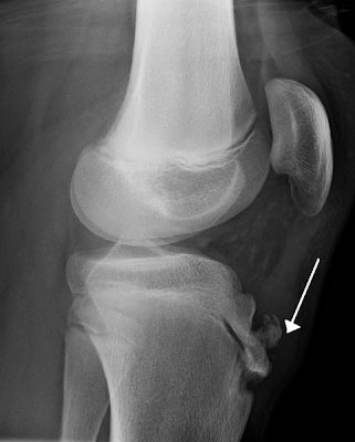
X-ray Osgood-Schlatter disease
Osgood Schlatters disease is a very common cause of knee pain in children and young athletes usually between the ages of 10 and 15. It occurs due to a period of rapid growth, combined with a high level of sporting activity.
* Normal x-ray findings do not exclude the disease, which is diagnosed clinically
* Radiographs have Limited role "Clinical diagnosis"...............
Imaging Findings
* Normal x-ray findings do not exclude the disease, which is diagnosed clinically
* Radiographs have Limited role "Clinical diagnosis"...............
Lateral radiograph of the knee demonstrating fragmentation of the tibial tubercle with overlying soft tissue swelling.
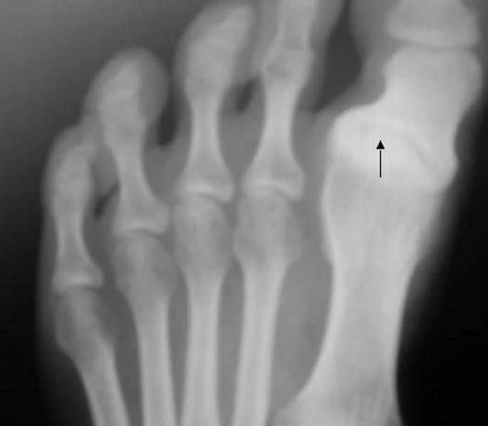
Hallux varus in X-ray

The condition has various degrees ..............
Acute pulmonary edema following surgery

- a. Left lower lobe pneumonia.
- b. Acute pulmonary edema.
- c. A large pneumothorax.
- d. A large pericardial effusion.
- e. A ruptured gastric ulcer.
Correct Answer: Acute pulmonary edema.
Explanation
There is diffuse airspace disease in both lungs causing almost complete opacification of both lungs. This came on suddenly and is characteristic of pulmonary edema. Other fluids can inhabit the airspaces such as blood or gastric aspirate, but they have different clinical stories and are less common than acute pulmonary edema. This patient had been hypotensive and was suffering from non-cardiogenic pulmonary edema. Vasopressors and diuretics were used in his treatment.
Scaphoid fractures overview

Anatomic snuffbox tenderness is a highly sensitive test for scaphoid fracture, whereas scaphoid compression pain and tenderness of the scaphoid tubercle tend to be more specific. Initial radiographs in patients suspected of having a scaphoid fracture should include anteroposterior, lateral, oblique, and scaphoid wrist views..........
READ MORE................>>

Subscribe to:
Posts (Atom)