Custom Search

Ultrasound image of a normal eye with lens

Since the eye is a fluid-filled structure, it provides a perfect acoustic window, producing images with excellent detail. The normal eye appears as a circular hypoechoic structure. The cornea is seen as a thin hypoechoic layer parallel to the eyelid.
The anterior chamber is filled with anechoic fluid and is bordered by the cornea, iris and anterior reflection of the lens capsule. The iris and ciliary body are seen as echogenic linear structures extending from the peripheral globe towards lens. The normal lens is anechoic. The normal vitreous chamber is filled with anechoic fluid. Vitreous is relatively echolucent in a young healthy eye.

Sonographically, the normal retina cannot be differentiated from the other choroidal layers. The evaluation of the retrobulbar area includes optic nerve, extraocular muscles and bony orbit. The optic nerve is visible posteriorly as a hypoechoic linear region radiating away from globe.
Normal ocular ultrasound

Ultrasound image of a normal eye with lens


Recurrent Pyogenic Cholangitis (RPC)


Recurrent pyogenic cholangitis,Cholangiohepatitis, is an uncommon disease in the western world Most of these cases are seen in Asian countries.
The etiology is unknown, although some of these patients have biliary parasites.
The disease is characterized by a recurrent syndrome of bacterial cholangitis that occurs in association with intrahepatic pigment stones and intrahepatic biliary obstruction.
-These patients are also at risk of developing biliary cirrhosis and cholangiocarcinoma.
-The left lobe is the most common location of the disease due to the delayed drainage of the left system.
This C.T. image shows a typical case of Recurrent pyogenic cholangitis
There is focal dilatation of the bile ducts in the left lobe with stones.

Below there is another example of recurrent pyogenic cholangitis.There is intrahepatic lithiasis with focal diatation.
A case like this is indistinguishable from focal Caroli disease with secundary stone formation.

Causes of lung collapse

Lung collapse usually occurs due to proximal occlusion of a bronchus, causing a loss of aeration. The remaining air is gradually absorbed, and the lung loses volume.

There are many causes for collapse, the most frequent are listed below:

* The commonest cause is a proximal stenosing bronchogenic carcinoma, which occludes a bronchus. Patients are middle aged or elderly, and almost always smokers.

* In a young adult or older child asthma is by far the commonest cause. Collapse occurs secondary to mucous plugging of the major airways, and responds to physiotherapy.
* In an infant consider an inhaled foreign body, such as a peanut.

* Retention of secretions is a frequent cause of post operative collapse.

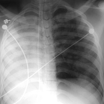
* In ventilated patients, including neonates, collapse may occur if the endotracheal tube is inserted too far, entering one main bronchus and occluding the other as shown below:
 Complete collapse of the right lung in this case was caused by an endotracheal tube that was advanced too far and entered the left mainstem bronchus. The heart is not seen as the mediastinum is shifted to the right. There is overinflation of the left lung, also contributing to the mediastinal shift.

Ultrasound showing Bilateral communicating hydrocele in neonate

The below ultrasound images show gross bilateral communicating hydrocele in a neonate, which was found to have Hydrops fetalis in utero. Note the presence of ascitic fluid with communication with the hydrocele (arrow in last figure below).


Benefits of Schüller view on showing Mastoid bone

Schüller's view (Runstrom) is a lateral view of the mastoid obtained with the sagittal
plane of the skull parallel to the film and with a 30° cephalocaudal angulation of the x-ray beam.

These 30° in Schüller's view displaces the arcuate eminence of the petrous bone downward and shows the antrum and the upper part of the attic.

Schüller's view also gives an excellent view of the extent of the pneumatization of the mastoid,the distribution and the degree of aeration of the air cells, the status of the trabecular pattern, and the position of the vertical portion of the lateral sinus.
Schüller view: Well-developed normally pneumatized mastoidal air cells can be observed in the picture on the left side (double arrow). In the picture on the right side, the mastoid cells (arrow) are obscured, and not air-containing, due to chronic otitis media.