Custom Search

Hutch diverticula of Bladder

A 73-year-old man presented to the emergency department after having intermittent fevers for 2 weeks. He had a history of recurrent urinary tract infections, parkinsonism, and a compression fracture at the L2 vertebra that was the result of a fall 2 years before presentation.
In addition, he had paraparesis and a neurogenic bladder, also subsequent to the fall. The results of physical and laboratory evaluation were notable for the identification of Pseudomonas aeruginosa in a blood culture and for a urinalysis showing more than 100 white cells per high-power field. He was treated with empirical antibiotics.

Intravenous urography showed no obstructive uropathy, but symmetric diverticula could be seen near both ureteral orifices (arrows). These lesions, known as Hutch diverticula, are usually congenital rather than occurring as a result of a neurogenic bladder or an infection or obstruction. They represented a new finding in this patient. Hutch diverticula are more commonly seen in men and boys and are usually unilateral and asymptomatic. After treatment with antibiotics, the patient's fever and pyuria subsided.
He declined any further evaluation or intervention. During the year after diagnosis, two more urinary tract infections developed.

Tuberculoma & Calcified tuberculoma

It appears Round or oval, sharply circumscribed nodule that is seldom more than 4 cm in diameter. Central calcification and “satelliteâ€‌ lesions are common, as is calcification of hilar lymph nodes.

Note that: Primarily involves the upper lobes (especially the right). The draining bronchus may show irregular thickening or even frank stenosis.


This X-ray shows :Single smooth, well-defined pulmonary nodule in the left upper lobe. In the absence of a central nidus of calcification, this appearance is indistinguishable from that of a malignancy.


(A) Frontal and (B) lateral views of the chest show a large left lung soft-tissue mass (arrows) containing dense central calcification.

Appearance of Cricopharyngeal achalasia in X-ray


It appeares as :Hemispherical or horizontal, shelflike protrusion on the posterior aspect of the esophagus at approximately the C5-C6 level.

Short Comment.......It is Failure of the upper esophageal sphincter to relax. Can result in dysphagia by obstructing the passage of a swallowed bolus. In severe disease, can cause aspiration and pneumonia.

Aortic Dissection

* 3:1 male to female predominance
* Over the age of 40
* Hemorrhage in the media (at vasa vasorum) leading to either
  1. Hemorrhage in the wall (less common)
  2. Hemorrhage separate media from adventitia

· Predisposing factors
o Hypertension (most commonly)
o Atherosclerosis
o Cystic medial necrosis
o Coarctation of the aorta
o Aortic stenosis
o S/P prosthetic aortic valve
o Trauma (rare)
o Pregnancy (rare)


· Aneurysm defined by size criteria
o In general, ascending aorta > 5 cm
o Descending aorta > 4 cm

· Vessels involved with dissection
o Any artery can be occluded
o Usually the right coronary and three arch vessels are involved with arch
aneurysms
o Right pulmonary artery and left-sided pulmonary veins may be occluded

· Types
o DeBakey Type I...............................................Involves entire aorta
o DeBakey Type II "Least common"...............Ascending aorta only
o DeBakey Type III "Most common"...............Descending aorta only
o Stanford Type A................................................Ascending aorta involved
----- Over half develop aortic regurgitation
o Stanford Type B.................................................Ascending aorta NOT involved
· Most dissections arise either just distal to the aortic valve or just distal to aortic isthmus


·Clinical
o Sharp, tearing, intractable chest pain

o Murmur or bruit of aortic regurgitation

o Previously hypertensive, now possible shock

o Asymmetric peripheral pulses

o Pulmonary edema


· Imaging Findings
o Chest films
  • - Mediastinal widening
  • - Left paraspinal stripe
  • - Displacement of intimal calcifications
  • - Apical pleural cap
  • - Left pleural effusion
  • - Displacement of endotracheal tube or nasogastric tube

o MRI
  • - Intimal flap
  • - Slow flow or clot in false lumen

o CT
  • - Intimal flap
  • - Displacement of intimal calcification
  • - Differential contrast enhancement of true versus false lumen

CT of abdominal aorta shows intimal flap (dark line -red arrow)
with true lumen anteriorly and false lumen posteriorly

o Angiography
  • - Intimal flap
  • - Double lumen
  • - Compression of true lumen by false channel
  • - Increase in aortic wall thickness > 10 mm
  • - Obstruction of branch vessels

Diagnosing of spinal cord compression

An accurate history and physical examination is essential in diagnosing spinal cord compression. The neurologic examination often identifies the suspicious areas allowing for improved accuracy in imaging the appropriate affected areas of the spine. Patients with spinal cord compression often have abnormalities on plain radiographs of the spine. The abnormalities encountered may include bony erosion and pedicle loss, partial or complete vertebral collapse, and paraspinous soft tissue masses. However, normal spine films do not exclude epidural metastasis.

MR tomography of the spine is the best method for evaluating epidural spinal cord compression. Gadolinium enhancement may be utilized when there is suspicion of cord compression due to epidural abscess. Gadolinium will enhance inflamed tissues and will define anatomic margins. Myelography requires an experienced physician and accompanied by CT may be performed with minimum patient discomfort. However, when metastatic disease completely blocks the spinal cord, myelography will not allow definition of the upper margin of tumor involvement.

Spinal cord compression. This sagittal magnetic resonance imaging scan of the spine demonstrates compression of the lower portion of the spinal cord by tumor (arrow head). Metastatic disease in the vertebral body is also seen.

Age-related Maculopathy with Angiogram

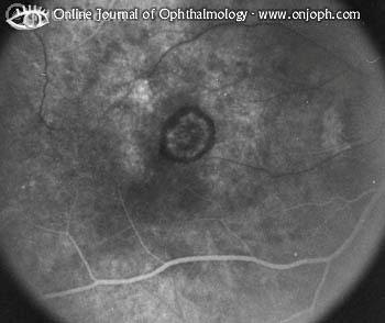
This photo shows age-related Maculopathy AMD, Classic Choroidal Neovasculariz.

Note that in an area of slight depigmentation (presumably a previous RPE detachment) is a darkly pigmented spot surrounded by a slim ring of subretinal blood.


With Angiogram:
#In the arterial phase the cartwheel of new choroidal vessels becomes immediately visible (together with the choroidal flush). A slight window defect is in the area of hypopigmentation.

#In mid-angiogram the neovascularization becomes prominent. The ring of surrounding blood blocks fluorescence.

#Late angiogram with diffuse leakage from the new vessels even into some of the blood.

A case of Giant Left Atrium


An 83-year-old woman with long-standing atrial fibrillation who had previously undergone atrioventricular nodal ablation and pacemaker placement presented with symptoms of progressive heart failure.

Physical examination
was notable for elevated jugular venous pressure, precordial lift, a grade 2/6 holosystolic murmur at the sternal border and apex, hepatomegaly, ascites, and severe lower-extremity edema.

Laboratory evaluation revealed a creatinine level of 1.4 mg per deciliter (124 µmol per liter), an albumin level of 3.6 g per deciliter, and a brain natriuretic peptide level of 526 pg per milliliter (normal range, 0 to 100 pg per milliliter); liver function was normal.

Chest radiography
(Panel A) revealed cardiomegaly (cardiothoracic ratio, 0.86), splaying of the carina, and an elevated left main bronchus (arrows).
An echocardiogram showed massive biatrial enlargement (left larger than right), normal ventricular size and function, and moderate mitral and tricuspid regurgitation (Panel B; LA denotes left atrium, LV left ventricle, RA right atrium, and RV right ventricle).
An esophagogram (Panel C) obtained to evaluate dysphagia for solid food revealed a prominent impression of the left atrium on the esophagus (E), without evidence of obstruction.
The patient was discharged home on medical management after prolonged diuresis.

velvet palm as a paraneoplastic sign

A 54-year-old woman who had a 34-pack-year smoking history presented with a 5-month history of changes in palmar skin texture associated with a 15-kg weight loss and left hip pain waking her at night.

On physical examination, cachexia was noted, along with a rugated, velvety appearance of the palms (Panel A), acanthosis nigricans, and left hip pain on rotation.

Laboratory evaluation
revealed neutrophil leukocytosis (white-cell count, 42,300 per cubic millimeter), thrombocytosis (platelet count, 1.17 million per cubic millimeter), and hypercalcemia (calcium corrected for albumin, 4.5 mmol per liter [18 mg per deciliter]).

Chest computed tomography (CT) revealed nodules (Panel B, arrow), and bone scintigraphy showed increased uptake in the left hip. Both these radiographic findings suggest metastasis. CT-guided fine-needle aspiration of a lung nodule revealed cells consistent with non–small-cell carcinoma of the lung.

The palmar changes represent a cutaneous paraneoplastic sign known as tripe or velvet palm. It is highly associated with cancer (94% in some reports), most frequently with gastric adenocarcinoma or bronchogenic carcinoma. The patient was referred to oncology for chemotherapy but died a few weeks later of metastatic lung cancer.

Coal workers pneumoconiosis - stage II


This chest x-ray shows stage II coal worker's pneumoconiosis (CWP). There are diffuse, small light areas on both sides of the lungs. Other diseases that may explain these x-ray findings include simple silicosis, disseminated tuberculosis, metastatic lung cancer, and other diffuse, infiltrative pulmonary diseases.

Vocal cord Paralysis

This can be caused for a variety of reasons, including tumors in the neck or trauma. Once or even both vocal cords can be affected. Surgery may be performed to readjust the vocal cords to restore normal speaking and breathing.


MRI scan demonstrating mass in neck causing paralyzed right vocal cord.

ABC's of Trauma :economic of chest radiograph

The chest radiograph is an economical and sensitive screening examination for the major injuries in the patient who has sustained blunt chest trauma. Just as the physician uses the ABC's to stabilize the critical ill patient (Airway, Breathing, Circulation), the radiologic ABC's prompt the radiologist to consider all of the critical injuries that may be sustained with blunt trauma. THE most critical injury is considered first.

  • Aortic Transection
  • Bronchial fracture
  • Cord injury (Thoracic spine)
  • Diaphragmatic rupture
  • Esophageal tear
  • Flail chest
  • Gas (subtle pneumothorax)
  • Heart (Cardiac injury)
  • Iatrogenic (Misplaced monitoring & support catheters)

Answer of the Case !!

What is the most likely diagnosis in the patient shown below?


a. Sprue

b. Scleroderma

c. Crohn's disease

d. Carcinoma of the colon

e. Duodenal hematoma


The Correct Answer: in 28April 10
Crohn's disease

Explanation
-----------

There is a long segment of terminal ileum which is narrowed with irregular margins. There are several ulcers or sacculations present. The location and appearance of this narrowed segment is characteristic of Crohn's Disease. This long narrowed segment would qualify as the "string-sign" of peristent spasm seen with that disease. Crohn's most often affects the terminal ileum and has a bimodal age distribution. Surgery is usually avoided because of the propensity for recurrence at the site of the neo-terminal ileum.