Hyperechoic amniotic fluid
These ultrasound images reveal markedly echogenic (hyperechoic) amniotic fluid. The fluid shows almost the same echogenicity as the placenta. Such appearances of the liquor amni on sonography are seen due presence of vernix caseosa (commonest cause), blood or meconium in the amniotic fluid.
Pneumonia in the Right middle lobe
The right middle lobe is bordered superiorly by the horizontal fissure, and medially by the right heart border. Any abnormality, which increases density of this lobe, may therefore obscure the right heart border, or be limited superiorly by the horizontal fissure.
This x-ray of Child with a cough and fever shows right middle lobe consolidation :
* The right heart border (right atrial edge) is obscured
* Consolidation is limited above by a crisp line, formed by the horizontal fissure "red arrow"
* The pathology must therefore involve the right middle lobe
* More extensive shadowing also involves the right and left peri-hilar regions
Diagnosis
* Pneumonia involving the right middle lobe
Subchorionic cyst lesion of the placenta
These ultrasound images show a cystic lesion in the placenta,located just below the placental surface. Few mobile echoes were seen within the lesion. No other abnormalities were seen in the placenta and the fetus. This finding is generally considered to be insignificant clinically. However some reports suggest an association with fetal IUGR (Intrauterine growth retardation).
It may be called:
subchorionic cyst of the placenta Or membranous cyst Or chorionic cyst.It is believed to be due to deposition of fibrin in the subchorionic region of the placenta.
CT scan of Blunt trauma to the spleen
• Spleen is the most commonly injured solid intra-abdominal organ and the Blunt trauma is the most common cause.
• Often (40%) associated with lower rib fractures and left renal injury.
• In 20% of patients with left rib fractures, there is a concomitant splenic injury.
• 25% of patients with left renal injuries also have splenic injuries.
• Damage ranges from subcapsular haematoma to total splenic laceration, potentially leading to exsanguination.
Rupture of the spleen. This CT shows Rupture of the anterior half of the spleen caused by blunt trauma in falling from a horse.Haemorrhage is seen within the splenic bed (arrow) along with free blood around the liver (arrowhead).
Splenic laceration (arrow).
Appearance of Hydronephrosis in ultrasound
Hydronephrosis refers to a kidney with a dilated pelvis and collecting system. It can be caused by obstruction of the ureters or bladder outlet. Hydronephrosis can also result from reflux (retrograde leakage of urine from the bladder up the ureters to the renal pelvis. Rarely, some children have hydronephrosis without either obstruction or reflux. This is thought to result form abnormal smooth muscles of the renal pelvis or ureter causing ectasia.
This ultrasound image demonstrates dilated renal calyces indicative of hydronephrosis. Chronic reflux uropathy can lead to hydronephrosis which can result in renal dysfunction as the calyces dilate and compress the renal parenchyma.
*Black arrow = renal capsule
*Black arrowhead = sinus fat
*White arrow = dilated calyx
*White arrowhead = renal cortex
This longitudinal ultrasound shows a kidney with moderate hydronephrosis. The parenchyma is relatively normal in thickness. The dilation of the collecting system extends from the renal pelvis to the calyces. This is a grade III hydronephrosis.
Ultrasound advantage in Renal calculi investigations
Renal calculi are commonly arise within the collecting renal system. Plain radiographs and intravenous urography are the traditional investigations for renal colic. Ultrasound has the advantage of demonstrating non-opaque calculi and hydronephrosis in comparison with the plain radiographs. Ultrasound has the potential of early diagnosis as compared with intravenous urography.
Hydronephrosis and hydrourether may also be present. Tiny calculi may not cause distal shadowing if they are smaller than the focal zone of the transducer. When there is no shadowing, it may be difficult to distinguish small calculi from echogenic foci caused by fat or other echogenic reflectors within the renal sinus.
Staghorn calculi often appear as multiple, disconnected calculi within the collecting system . Sonography generally underestimates the extent and size of stones in patients with staghorn and other large calculi. The presence of staghorn calculi may make it difficult to diagnose underlying hydronephrosis, because of strong acoustic shadowing from calculi.
Appearance of Renal stones by Ultrasound :
Most renal calculi (about 80%) are calcified. Sonographically, stones usually appear as a hyperechoic foci with distal acoustic shadowing . Gas may cause a similar appearance but may have a "dirty" shadow that is not as sharply defined as would occur with a calculus. There may be associated mucosal edema if the stone is impacted or if there is secondary inflammation or infection. Hydronephrosis and hydrourether may also be present. Tiny calculi may not cause distal shadowing if they are smaller than the focal zone of the transducer. When there is no shadowing, it may be difficult to distinguish small calculi from echogenic foci caused by fat or other echogenic reflectors within the renal sinus.
 |
| Staghorn calculi |
Vertebral artery aneurysm
This Vertebral artery aneurysm or tortuosity causes enlarged cervical intervertebral foramen.
Erosion is caused by pulsatile flow as the vertebral artery passes through the foramina transversaria of the upper six cervical vertebrae between its origin from the subclavian artery and its entrance into the cranial vault through the foramen magnum.
Erosion is caused by pulsatile flow as the vertebral artery passes through the foramina transversaria of the upper six cervical vertebrae between its origin from the subclavian artery and its entrance into the cranial vault through the foramen magnum.
Tortuous vertebral artery. (A) Frontal tomogram shows the enlarged foramen (arrows). (B) Arteriogram shows the tortuous vertebral artery (arrow) entering the enlarged foramen.
Optic nerve glioma; the most common cause of optic nerve thickening
Optic nerve glioma appears as diffuse enlargement of the left optic nerve (arrows) in an 8-year-old girl.
Optic nerve glioma is the most common cause of optic nerve enlargement. Typically causes uniform thickening of the nerve with mild undulation or lobulation. In children (especially preadolescent girls), optic nerve gliomas are usually hamartomas that spontaneously stop enlarging and require no treatment. In older patients, however, these gliomas may have a progressive malignant course despite surgical or radiation therapy. Optic nerve gliomas are a common manifestation of neurofibromatosis (typically low-grade lesions that act more like hyperplasia than neoplasms).
Optic nerve glioma. (A) Sagittal and (B) coronal T1-weighted MR scans show involvement of the chiasm and left optic nerve.
Radiological abnormality in case of tension pneumothorax
A 23-y man with well past health, presented with sudden onset of left sided chest pain and shortness of breath, This pain was sharp in nature and more severe on inspiration. Physical examination showed decreased air entry in the left upper chest which was hyperresonant on percussion. Laboratory investigations were essentially normal. A CXR was performed for further evaluation .
- Hyperlucent zone devoid of vascular marking in periphery of left hemithorax.
- Shift of midline to the right.
(2) So; the most likely diagnosis is left tension pneumothorax
Note the radiological features of tension pneumothorax seen in this
patient include:
- Contralateral mediastinal shift
- Depression of ipsilateral hemidiaphragm
- Compressive atelectasis of adjacent normal lung
All of the above radiological signs indicate the presence of significant increased intra-thoracic pressure in tension pneumothorax which necessitates urgent treatment.
* The absence of vascular markings in the periphery of the left hemithorax is due to air in the pleural cavity and not in the lung.
* Role of imaging in patients with pneumothorax:
1. Confi rm the clinical diagnosis
2. Assess extent of pneumothorax
3. Detect signs of tension pneumothorax
4. Follow-up examination to monitor resolution of pneumothorax after drainage
Large left pneumothorax with mediastinal shift to the right. Note the collapsed left lung (arrows) and the hyperlucent left hemithorax.
(1) The radiological abnormality that can be identified :- Hyperlucent zone devoid of vascular marking in periphery of left hemithorax.
- Shift of midline to the right.
(2) So; the most likely diagnosis is left tension pneumothorax
Note the radiological features of tension pneumothorax seen in this
patient include:
- Contralateral mediastinal shift
- Depression of ipsilateral hemidiaphragm
- Compressive atelectasis of adjacent normal lung
All of the above radiological signs indicate the presence of significant increased intra-thoracic pressure in tension pneumothorax which necessitates urgent treatment.
Notes:
* The diagnosis of pneumothorax is confirmed by erect chest radiograph in full expiration. * The absence of vascular markings in the periphery of the left hemithorax is due to air in the pleural cavity and not in the lung.
* Role of imaging in patients with pneumothorax:
1. Confi rm the clinical diagnosis
2. Assess extent of pneumothorax
3. Detect signs of tension pneumothorax
4. Follow-up examination to monitor resolution of pneumothorax after drainage
Continuous diaphragm sign of pneumomediastinum and pneumoperitoneum
 Definition of continuous diaphragm sign of pneumomediastinum:
Continuous diaphragm sign is seen when The entire diaphragm is visualized from one side to the other because air in the mediastinum outlines the central portion which is usually obscured by the heart and mediastinal soft tissue structures that are in contact with the diaphragm.
Also;continuous diaphragm sign of pneumoperitoneum
One of the manifestation of massive pneumoperitoneum is the continuous diaphragm sign. Where there is sufficient air beneath the diaphragm, the continuous nature of the diaphragm is demonstrated. Note that the left and right hemidiaphragms contrasted by the free gas appear as a continuous structure.
continuous diaphragm sign of pneumoperitoneum
Lung collapse due to bronchogenic carcinoma with ‘Golden S sign’
A 70-year-old chronic smoker presented with haemoptysis and weight loss for 2 months. He had no fever, chills or rigor and a physical examination of both hands showed fi nger clubbing. There was decreased chest wall expansion and air entry over right upper chest. Laboratory investigations were essentially unremarkable and WCC was within normal limits. A CXR was performed for further evaluation .
- Focal convex bulge at the apex of the abnormality.
- Hyperinflation of the right lower lobe.
- Elevated right hemidiaphragm.
radiological diagnosis: Lung collapse
- The lack of air within collapsed right upper lobe accounts for the increase in radiographic density.The well-demarcated lateral border represents the elevated horizontal fissure.
- The focal bulge at the apex of the collapsed right upper lobe corresponds to the centrally located bronchogenic carcinoma causing the lobar collapse. The combined radiologic appearance on frontal radiograph is known as ‘Golden S sign’.
- The hyperinflation and elevated right hemidiaphragm are due to volume loss.
1. Crowding of ribs in right upper chest wall – due to underlying lung volume loss
2. Tracheal deviation to the right – due to traction from the collapsed lung
* Recognition of lobar collapse is important, especially in elderly patients, as this may be the only radiologic feature of primary lung carcinoma. Further evaluation by sputum cytology, bronchoscopy or CT scan is necessary.
The ‘Golden S sign’ – collapse of the right upper lobe with a well demarcated lateral border formed by the elevated horizontal fissure (arrows), and a focal convex bulge at the apex due to the centrally located bronchogenic carcinoma (arrowheads).
The abnormalities that can you see on this CXR :
- Opacity with a sharp well-demarcated lateral border (arrows) in right upper zone with lack of air within the abnormality. - Focal convex bulge at the apex of the abnormality.
- Hyperinflation of the right lower lobe.
- Elevated right hemidiaphragm.
radiological diagnosis: Lung collapse
- The lack of air within collapsed right upper lobe accounts for the increase in radiographic density.The well-demarcated lateral border represents the elevated horizontal fissure.
- The focal bulge at the apex of the collapsed right upper lobe corresponds to the centrally located bronchogenic carcinoma causing the lobar collapse. The combined radiologic appearance on frontal radiograph is known as ‘Golden S sign’.
- The hyperinflation and elevated right hemidiaphragm are due to volume loss.
Notes:
* Other radiologic features of right upper lobe collapse not seen on this chest radiograph include:1. Crowding of ribs in right upper chest wall – due to underlying lung volume loss
2. Tracheal deviation to the right – due to traction from the collapsed lung
* Recognition of lobar collapse is important, especially in elderly patients, as this may be the only radiologic feature of primary lung carcinoma. Further evaluation by sputum cytology, bronchoscopy or CT scan is necessary.
A Pancreatic mass or just papillary process of the caudate lobe of liver
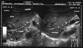
This patient presented with abdominal discomfort and underwent sonography of the abdomen. Ultrasound images show a rounded mass in the region of the pancreatic head and isthmus. It shows the same echogenicity as the liver (photos 1 and 2). This suggested the possibility of a pancreatic mass, possibly malignant.
1-
2-
However, images 3 and 4, reveal a different diagnosis- the possible "mass" appears to be an extension of the caudate lobe of the liver. These ultrasound images are diagnostic of "papillary process of the caudate lobe of liver."This normal variant may thus mimic pancreatic or preaortic lymph node masses.
3-
4-
Images courtesy of Dr. Ravi Kadasne, UAE.
What is Serendipity View or Rockwood view? And how to get?
Serendipity View "some times called Rockwood view" obtained to demonstrates sternoclavicular joints and medial 1/3 of the clavicles for fractures or dislocation.
1-The beam is angled 40 deg. off verticle centered on the sternum.
2-Tube to cassette distance is 60 deg for adults and 40 deg for child.
$$ Note that CT scan best evaluates the sternoclavicular joint
Technique of Serendipity View :
Non rigid 11 x 14 inch cassette is placed under the upper chest, shoulders, and neck of a supine patient.1-The beam is angled 40 deg. off verticle centered on the sternum.
2-Tube to cassette distance is 60 deg for adults and 40 deg for child.
Why we need it ?
As Medial clavicular fractures and SC joint injuries may be difficult to appreciate on standard views because of the overlap of the clavicle with the sternum and the first rib.Normal Rockwood (Serendipity) view of the sternoclavicular (SC) joint.
$$ Note that CT scan best evaluates the sternoclavicular joint
Radiological features of Bezoar
Bezoar is an intestinal mass caused by the accumulation of ingested
material which Can lead to obstruction or ulceration.
Bezoar Types :
• A phytobezoar is formed from poorly digested plant fibre.
• A trichobezoar is formed from ingested hair, almost always in females.
• May demonstrate bowel obstruction.
• Barium may flow into crevices within thebezoar.
CT:
• This may demonstrate a low-density mass containing pockets of air.
• As on barium studies, oral contrast may intersperse with the mass
though gaps between the ingested materials.
material which Can lead to obstruction or ulceration.
Bezoar Types :
• A phytobezoar is formed from poorly digested plant fibre.
• A trichobezoar is formed from ingested hair, almost always in females.
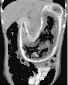
Trichobezoar. Large ‘hair ball’ mass completely filling the stomach (arrow).
Radiological features :
AXR:
• A mass may be seen within the stomach.• May demonstrate bowel obstruction.
Barium studies:
• May demonstrate an intraluminal filling defect that does not have a fixed site of attachment to the bowel wall.• Barium may flow into crevices within thebezoar.
CT:
• This may demonstrate a low-density mass containing pockets of air.
• As on barium studies, oral contrast may intersperse with the mass
though gaps between the ingested materials.
Trichobezoar (same patient) in coronal CT reformat.Oral contrast is seen outlining the huge trichobezoar.
Psoriatic arthritis in the sacroiliac joint
Psoriatic arthritis in the sacroiliac joint usually appears with bilateral, asymmetric distribution , though bilateral, symmetric abnormalities are frequent and even unilateral involvement may occur.
The radiographic changes include erosions and sclerosis, predominantly affecting the ilium, and widening of the articular space. Although joint space narrowing and bony ankylosis can occur, this is much less frequent than in classic ankylosing spondylitis. A prominent finding may be blurring and eburnation of apposing sacral and iliac surfaces above the true joint in the region of the interosseous ligament.
Psoriatic arthritis in the sacroiliac joint
The radiographic changes include erosions and sclerosis, predominantly affecting the ilium, and widening of the articular space. Although joint space narrowing and bony ankylosis can occur, this is much less frequent than in classic ankylosing spondylitis. A prominent finding may be blurring and eburnation of apposing sacral and iliac surfaces above the true joint in the region of the interosseous ligament.
Psoriatic arthritis. Bilateral, though somewhat asymmetric, narrowing of the sacroiliac joints.
Voluminous inflammatory exudate due to Klebsiella pneumonia
Pneumonia due to Klebsiella infection Tends to form a voluminous inflammatory exudate that produces a homogeneous parenchymal consolidation (containing an air bronchogram) and bulging of an interlobar fissure. High frequency of abscess and cavity formation (rare in pneumococcal pneumonia).
Klebsiella pneumonia. Downward bulging of the minor fissure (arrow) due to massive enlargement of the right upper lobe with inflammatory exudate.
Aortic arch in MRA and its Variations
The aortic arch is the direct continuation of the ascending aorta. Its origin is defined as sternomanubrial joint. It terminates at the lower border of T4, when it becomes the descending aorta.
Branches :
Three main branches typically originate from the upward convexity of the arch. In order from proximal to distal these are:
1- (right) brachiocephalic artery (or innominate artery) which goes on to divided into the right subclavian and right common carotid arteries.
2- left common carotid artery
3- left subclavian artery
Variations
There are three common variations to the branching pattern:
1. normal (as described above) that seen in ~ 70% of patients.
2. bovine arch : common origin of brachiocephalic and left common carotid artery - seen in approximately 15% of patients (more common in blacks)
3. left common carotid has its origin from the brachiocephalic artery proper, rather than from a common trunk : seen in approximately 10% of patients (also more common in blacks)
* thyroidea ima artery, usually between the brachiocephalic and left common carotid
* left vertebral artery, usually between the left common carotid and the left subclavian arteries.
* rarely the right subclavian and right common carotid arise independently.
Branches :
Three main branches typically originate from the upward convexity of the arch. In order from proximal to distal these are:
1- (right) brachiocephalic artery (or innominate artery) which goes on to divided into the right subclavian and right common carotid arteries.
2- left common carotid artery
3- left subclavian artery
Variations
There are three common variations to the branching pattern:
1. normal (as described above) that seen in ~ 70% of patients.
2. bovine arch : common origin of brachiocephalic and left common carotid artery - seen in approximately 15% of patients (more common in blacks)
3. left common carotid has its origin from the brachiocephalic artery proper, rather than from a common trunk : seen in approximately 10% of patients (also more common in blacks)
Left ICA coming off the right brachiocephalic.
Additionally there may be additional branches that arise directly from the arch. These include* thyroidea ima artery, usually between the brachiocephalic and left common carotid
* left vertebral artery, usually between the left common carotid and the left subclavian arteries.
* rarely the right subclavian and right common carotid arise independently.
Simple bone cyst in x-ray
Simple bone cyst appears in x-ray as Expansile lucent lesion that is sharply demarcated from adjacent normal bone. May contain thin septa (scalloping of underlying cortex) that produce a multiloculated appearance. Tends to have an oval configuration with its long axis parallel to that of the host bone.
Simple bone cyst in the proximal humerus. The cyst has an oval configuration, with its long axis parallel to that of the host bone. Note the thin septa that produce a multiloculated appearance.
Notes :
Simple bone cyst is a True fluid-filled cyst with a wall of fibrous tissue. Begins adjacent to the epiphyseal plate and appears to migrate down the shaft (in reality, the epiphysis has migrated away from the cyst). Bone cysts arise in children and adolescents and most commonly involve the proximal humerus and femur. Often presents as a pathologic fracture that may show the fallen fragment sign (fragments of cortical bone are free to fall to the dependent portion of the fluid-filled cyst, unlike a bone tumor that has a firm tissue consistency).
Typical flow of cerebrospinal fluid (CSF) in MRI image
Sagittal T1-weighted image of the normal brain showingtypical flow of cerebrospinal fluid (CSF).
In T1-weighted magnetic resonance imaging sequences, CSF is black. From the paired lateral ventricles (LV), CSF passes through the paired interventricular foramina of Monro (yellow arrow) into the single midline third ventricle (TV). CSF then flows down the single midline aqueduct of Sylvius (a channel shaped like a toothpick and slender in all diameters; green arrow) into the single midline fourth ventricle (FV). CSF leaves the ventricular system through the two lateral foramina of Luschka and the midline foramen of Magendie. Here, CSF is shown exiting through the foramen of Magendie (blue arrow) and entering the cisterna magna (CM). Within the subarachnoid space (SAS), CSF flows over the convexities of the brain and the folia of the cerebellum, and around the brainstem (curved arrows). From the CM, CSF also courses inferiorly to surround the spinal cord (orange arrow).
Chest x-ray , A case of pneumonia
Video will describe how pneumonia may look like on a chest x-ray. Subtle pneumonia.
How to read the chest x ray :Concepts and Quality
Basic concepts of radiology and indicators of quality of the chest x ray.
Diagram of Anterior anatomical relations of both kidneys
The kidneys are retroperitoneal organs that are located in the perirenal retroperitoneal space with a longitudinal diameter of 10–12 cm and a latero-lateral diameter of 3–5 cm and a weight of 250–270 g.
In the supine position, the medial border of the normal kidney is much more anterior than the lateral border, The upper pole of each kidney is situated more posteriorly than the lower pole.
The right kidney, anteriorly :
has a relation with the inferior surface of the liver with peritoneal interposition,and with the second portion of the duodenum without any peritoneal interposition since the second portion of the duodenum is retroperitoneal .
The left kidney, anteriorly :
has a relation with the pancreatic tail, the spleen, the stomach, the ligament of Treitz and small bowel, and with the left colic lexure and left colon . Over the left kidney, there are two important peritoneal relections, one vertical corresponding to the spleno-renal ligament (connected to
the gastro-diaphragmatic and gastrosplenic ligaments) and one horizontal corresponding to the transverse mesocolon.


