How to identify Hematoma in Mammography ??

In this article We will show WELL-CIRCUMSCRIBED and ILL-DEFINED HEMATOMA as seen in Mammogaphy...

WELL-CIRCUMSCRIBED HEMATOMA:
Most commonly caused by blunt or surgical trauma, although hematomas may develop in patients who are anticoagulated or have clotting abnormalities. The combination of hemorrhage and edema more commonly results in an ill-defined mass or a diffuse area of increased density. Although the mammographic findings simulate carcinoma, a history of trauma suggests a conservative approach. Follow-up examinations show gradual decrease in size or even disappearance of the lesion. An organized hematoma may occasionally persist as a more sharply defined mass.

Imaging Findings:
Medium to high-density mass, often having slightly irregular margins. Overlying skin edema is usually present in the acute stage if the hematoma is secondary to trauma.
Hematoma. (A) Mammogram of a firm, palpable mass that arose at a recent biopsy site shows a dense lesion associated with skin thickening (arrows). (B) Three months later, there has been almost complete resolution of the hematoma with only minimal residual architectural distortion (arrows).


ILL-DEFINED HEMATOMA
Overlying skin thickening from edema and bruising may simulate carcinoma. Hematomas tend to resolve within 3 to 4 weeks.


Imaging Findings:
May appear as an ill-defined lesion (more commonly a relatively well-defined mass or a diffuse increase in density).

Hematoma. Ill-defined area of increased density (arrows) in the area of a lumpectomy performed 2 weeks previously.

Thoracic ultrasound for pneumothorax

instructional video discussing the use of thoracic ultrasound for pneumothorax

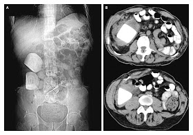
Calcifications in a Continent Urinary Diversion


A 37-year-old man with a history of spina bifida presented with an audible knocking sound coming from his abdomen during ambulation. Fifteen years earlier he had undergone a cystectomy with a continent cutaneous urinary diversion (Indiana pouch) for a neurogenic bladder. He had been poorly compliant with the catheterization and irrigation regimen involving the pouch.
Physical examination revealed a well-healed, functional stoma and a palpable mass in the right side of the abdomen. Urinalysis revealed a urinary pH of 9.0, with numerous white and red cells, and urine culture grew Proteus mirabilis.

Plain abdominal radiography
revealed two large multilaminar calcifications in the right side of the abdomen (Panel A). Computed tomography of the abdomen and pelvis confirmed the presence of two large calculi within the Indiana pouch (Panel B). The patient underwent cystolithotomy without incident. At a follow-up visit 1 year after the procedure, he was found to have been compliant with his catheterization and irrigation regimen and remained free of stones.

Continent urinary diversions may be associated with hypercalciuria, hyperphosphaturia, hyperoxaluria, and hypocitraturia, all of which may predispose patients to pouch urolithiasis. Infections with organisms that break down urea can result in an alkaline environment, which can in turn give rise to struvite (magnesium ammonium phosphate) stones. Establishment of a formal catheterization and irrigation protocol for continent urinary diversions may reduce the risk of urolithiasis.

Rectal bezoars in children

A bezoar is a solid mass formed in the gastrointestinal tract as a result of the consumption of indigestible or poorly digestible substances. Bezoars most commonly form in the stomach, but can occur in the small intestine and, rarely, in the colon or rectum. They are usually classified according to the material of which they are derived, for example, trichobezoars (hair), lactobezoars (milk curd), phytobezoars (plant fibre) and medication bezoars. The term originates from the Arabic term badzehar, meaning "antidote," a reference to historical medicinal use of these concretions found in animal gastrointestinal tracts in the treatment of human poisonings.

See this case of a previously well 8 year-old boy was brought to the emergency department with a three-day history of constipation. He had consumed three handfuls of flavoured unshelled sunflower seeds four days previously. His mother had unsuccessfully tried several sodium biphosphate and sodium phosphate enemas at home, as well as the use of bran to aid defecation. On examination, the patient’s abdomen was soft and nontender with normal bowel sounds. A bolus of stool mixed with unshelled sunflower seeds was visible at the anus.

A radiograph of the abdomen showed stool extending from the descending colon down to the rectum, which contained a large mass . Oral phosphate soda and a mineral oil enema were ineffective. The patient was taken to the operating room for digital and instrumental extraction under general anesthesia. He was monitored in hospital for 24 hours and discharged when he was taking liquids and passing soft stool.

Although small rectal bezoars may pass spontaneously, options for the care of patients presenting to clincians with symptomatic rectal bezoars include the use of enemas and extractions with conscious sedation or general anesthesia. Formation of bezoars from sunflower seeds may be more likely in children or preteens who are unable or unwilling to spit out the shells, particularly with flavoured shells.

Interventional Radiology: The Specialty, Patients, Procedure

Interventional radiology: the cutting edge of medicine without the cutting

“Cotton Wool” Appearance of Paget's Disease

A 63-year-old man presented with a long-standing history of sinusitis and 3 weeks of frontal headache. The physical examination was unremarkable.
The alkaline phosphatase level was elevated at 434 IU per liter (upper limit of the normal range, 129). The serum calcium level was within normal limits.

Radiography of the skull (Panel A) showed thickening of the outer and inner tables of the cranial bones, widening of the diploĆ«, and a “cotton wool” appearance caused by irregular areas of sclerosis (arrows). Computed tomography of the skull (Panel B) confirmed bony expansion, cortical bone thickening, and irregular areas of sclerosis (arrows). These imaging findings reflect the mixed osteolytic and osteoblastic phases of Paget's disease, resulting in accelerated bone turnover with bone deposition and expansion.
The patient was treated with alendronate, which resulted in improvement in frontal headache.

Diplopia in a Patient with HIV Infection


A 25-year-old man with human immunodeficiency virus (HIV) infection who was receiving highly active antiretroviral therapy presented with a 1-week history of diplopia and headache. The CD4 count was 218 cells per cubic millimeter, and the viral load was 50,000 copies per milliliter.
The neurologic examination revealed an inability to abduct the right eye with horizontal gaze, a finding that was consistent with an isolated right abducens nerve palsy (rightward gaze, Panel A; leftward gaze, Panel B). The examination of other cranial nerves was normal. The remainder of the motor and sensory examination was within normal limits.

The patient reported having had low back pain and constipation for the previous week. There was no history of bowel or bladder incontinence. A gadolinium-enhanced magnetic resonance image of the brain showed a minimally enhancing mass filling and expanding the right cavernous sinus (Panel C, arrow). Lumbar-spine imaging showed a mass with similar radiographic characteristics involving the ventral epidural compartment. Biopsy of the spinal lesion revealed diffuse large-B-cell lymphoma. A chemotherapeutic regimen of cyclophosphamide, doxorubicin, vincristine, and prednisolone, along with the monoclonal antibody rituximab (R-CHOP), was started. Progressive leg weakness from spinal involvement developed, and the patient had a poor response to corticosteroids and radiation therapy and died 3 months later.

CT & MRI in ICH

Magnetic Resonance Imaging and Computed Tomography in Emergency Assessment of Patients with Suspected Acute Stroke: A Prospective Comparison.

BACKGROUND: MRI is believed to be more sensitive than CT scanning for imaging ischemic stroke, but is considered insufficiently sensitive for acute hemorrhagic stroke.

METHODS: In this single-center study at a suburban hospital in Bethesda, MD, and coordinated at the National Institutes of Health, 356 patients with suspected acute strokes underwent MRI and CT scanning of the brain. The imaging studies were later reviewed by a panel of two neuroradiologists and two stroke neurologists blinded to the patients' clinical findings.

RESULTS: The final diagnosis was acute ischemic stroke in 190 patients (53%), acute intracranial hemorrhage in 27 (8%) and transient ischemic attack in 14 percent. Twenty-five percent of the patients were not ultimately diagnosed with cerebrovascular disease. The sensitivity of CT scanning was 26% for acute stroke overall, 16% for acute ischemic stroke and 89% for acute intracranial hemorrhage (corresponding specificities 98%, 98% and 100%, respectively). The sensitivity of MRI was 83% for acute stroke overall, 83% for acute ischemic stroke and 81% for acute intracranial hemorrhage (corresponding specificities, 97%, 96% and 100%, respectively). In the subgroup of patients imaged within three hours of symptom onset, sensitivities for acute stroke were 27% for CT scanning and 76% for MRI, and specificities were 100% and 96%, respectively.

CONCLUSIONS: Although the authors suggest that their findings are consistent with a superiority of MRI over CT scanning for the imaging of acute stroke, MRI missed more patients with acute hemorrhagic stroke, which could have significant implications when selecting patients for thrombolytic therapy.

What`s Caroli Disease and Caroli syndrome??

Caroli disease is one of non-obstructive biliary diseases that cause biliary dilatation,Once you have excluded obstruction, you have to think about nonobstructive biliary diseases as:

1- Caroli disease
2- Choledochal cyst
3- Recurrent pyogenic cholangitis
4- Primary sclerosing cholangitis

Caroli disease is an autosomal recessive disease secondary to malformation of the ductal plate .
It is associated with polycystic kidney disease, medullary sponge kidney and medullary cystic disease.
So looking at the kidneys can sometimes help you make this diagnosis.

In images below Notice the intrahepatic duct dilatation and the normal caliber of the choledochal duct (extrahepatic bile duct).
Saccular intrahepatic duct dilatation with normal sized choledochal duct in Caroli disease

Saccular bile duct dilatation in Caroli disease

**The hallmark of Caroli disease is intrahepatic duct dilatation.
The dilatation can be very large and saccular as seen in the image or it can be very linear.

The duct dilatation in Caroli disease is due to a congenital malformation of the ductal plate, which is the precursor of the intrahepatic bile ducts.we can see in this diagram the normal development of the ductal plate .
Embryologically each bile duct starts as a single layer of cells that surrounds a portal vein.after that this layer duplicates.
Portions of this double layer fuse and resorb leaving unfused portions that become the bile ducts.

So in the normal condition each portal vein is surrounded by interconnecting bile ducts .However if the patient has ductal plate malformation, the bile ducts are too numerous and they are ectatic (as seen below).
Whether or not we see this on imaging depends on which portion of the bile ducts is affected.
If the large ducts are involved, we see this as Caroli disease.
However if only the very small ducts are involved, the result is congenital hepatic fibrosis.
If all ducts are involved, then there is a combination of fibrosis and Caroli disease, which is also known as the Caroli syndrome.

Palpable Gall Bladder with CT


Palpable Gall Bladder: Cholangiocarcinoma has caused patient's gall bladder to become markedly dilated, and therefore,easily palpable (A).
Note skin is also jaundiced due to biliary tract obstruction. CT sagital image demonstrates dilated
gall bladder (B), dilated common bile duct (C), and dilated intrahepatic ducts (D). Courvoisier described the same
findings in patients with pancreatic cancer.

Chronic (total) renal infarction X-ray

Renal infarction due to acute renal artery occlusion. (A) An initial nephrotomogram demonstrates a thin cortical rim surrounding the right kidney (arrows), reflecting viable renal cortex perfused by perforating collateral vessels from the renal capsule. (B) Four months later, a repeat nephrotomogram shows a marked decrease in the size of the atrophic right kidney (arrowheads).

Imaging Findings :
Global shrinkage of the kidney with absent opacification. There may be a peripheral rim of opacified cortex during the nephrogram phase (probably reflects viable renal cortex perfused by perforating collateral vessels from the renal capsule).

NOTE : Renal occlusion is most commonly secondary to an embolism from the heart. A decrease in renal size is detectable within 2 weeks and reaches its maximum extent by 5 weeks. Compensatory enlargement of the contralateral kidney (in individuals young enough to provide this reserve).

Renal ischemia X-ray

Renal ischemia associated with hypertension. Diminished size of the right kidney (A) due to renal artery stenosis (B) (arrow).

In the category of Small, smooth kidney.It shows Unilateral delayed appearance and excretion of contrast material with subsequent hyperconcentration.
There may be ureteral notching (due to collateral arteries) and vascular calcification.

Note : Chronic ischemia (usually arteriosclerosis or fibromuscular hyperplasia) causes tubular atrophy and shrinkage of glomeruli. Often associated with hypertension, which is likely if the right kidney is at least 2 cm shorter than the left or if the left kidney is at least 1.5 cm shorter than the right. May be bilateral if there is generalized renal arteriosclerosis.

The Sandwich Sign

Transverse computed tomographic (CT) image obtained with oral and intravenous contrast material in a patient with mesenteric non-Hodgkin lymphoma (NHL). Image shows two large mesenteric lymphoma masses, which represent the halves of the sandwich bun (arrows), enveloping mesenteric fat and enhanced vessels.
Note that the enhanced small bowel lies outside of the sandwich. Extensive retroperitoneal lymphadenopathy (arrowheads) is present, as well.


Transverse CT image obtained with oral and intravenous contrast material in a patient who underwent left pelvic renal allograft transplantation 6 months earlier and now has mesenteric posttransplantation lymphoproliferative disorder (PTLD).
Mesenteric lymphoma mass (solid arrow) just to the right of the pelvic kidney (arrowhead) has produced a sandwich sign. Retroperitoneal lymphadenopathy (open arrows) is also seen.